Anatomy of the central complex
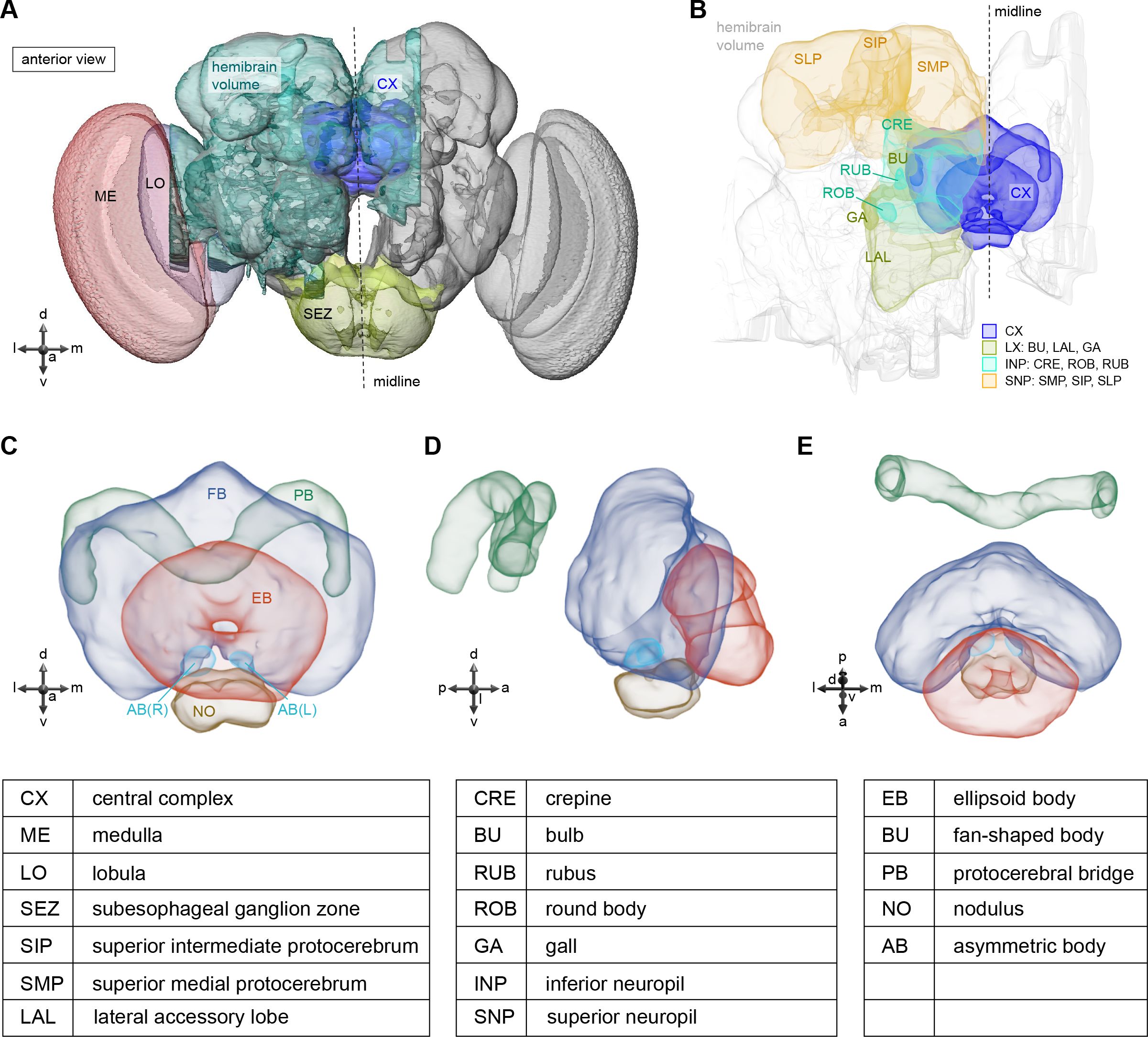
Anatomical overview of the adult central complex (CX) and its accessory neuropils, from Figure 1 of Hulse et al. (2021). Panel A places the CX within the hemibrain volume alongside the medulla (ME), lobula (LO) and subesophageal zone (SEZ). Panel B shows the immediate surrounds — superior protocerebrum (SLP, SIP, SMP), inferior neuropils (CRE, RUB, ROB) and lateral neuropils (BU, GA, LAL). Panels C–E give frontal, lateral, and dorsal views of the five core CX structures: ellipsoid body (EB), fan-shaped body (FB), protocerebral bridge (PB), paired noduli (NO), and the asymmetric body (AB). Click a labelled neuropil to open the corresponding term in Virtual Fly Brain.
Categories:
https://doi.org/10.7554/eLife.66039
Figure reproduced under CC-BY 4.0 from Hulse BK, Haberkern H, Franconville R, Turner-Evans DB, Takemura S, Wolff T, Noorman M, Dreher M, Dan C, Parekh R, Hermundstad AM, Rubin GM, Jayaraman V (2021). A connectome of the Drosophila central complex reveals network motifs suitable for flexible navigation and context-dependent action selection. eLife 10:e66039.
Feedback
Was this page helpful?
Glad to hear it! Please tell us how we can improve.
Sorry to hear that. Please tell us how we can improve.
Last modified May 12, 2026: Anatomy diagrams: cover label text below cell drawings (115a328)